
Oxidative stress: a new key player in T-cell lymphoma
CUTANEOUS T-CELL LYMPHOMA: Researchers from the LEO Foundation Skin Immunology Research Center (SIC) at the University of Copenhagen have uncovered a distinct redox imbalance in cutaneous T-cell lymphoma (CTCL) that leaves malignant T cells vulnerable to further oxidative stress and may point to new therapeutic opportunities.
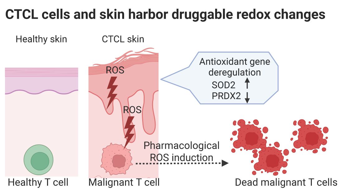
Reactive oxygen species (ROS) are natural by-products of cellular metabolism and play important roles in cell signaling. When ROS accumulate beyond the cell’s antioxidant capacity, however, they can damage DNA, proteins and lipid cell membranes. Although oxidative stress has been linked to inflammatory skin diseases and cancer, its role in CTCL has remained poorly understood.
In a new study, the researchers examined oxidative stress in CTCL skin lesions and in primary malignant T cells. They found increased oxidative damage in lesional mycosis fungoides skin together with a distinctive redox signature characterized by high levels of the antioxidant enzyme SOD2 and low levels of PRDX2.
The malignant T cells also displayed persistently elevated intracellular and mitochondrial ROS, indicating that they already operate close to the limit of their antioxidant capacity. When the researchers experimentally increased ROS further, malignant T cells were selectively killed. This effect could be prevented by the antioxidant N-acetyl cysteine, providing evidence that the cells are particularly sensitive to additional oxidative stress.
Importantly, the study shows that the consequences extend beyond the malignant cells themselves. Factors released by malignant T cells increased ROS in neighboring keratinocytes and reproduced the same SOD2-high/PRDX2-low redox profile. Activation by Staphylococcus aureus further amplified this effect, highlighting a potential connection between malignant T cells, bacteria and oxidative stress in the skin microenvironment.
The findings identify redox imbalance as a previously underexplored feature of CTCL and suggest that malignant T cells carry an intrinsic vulnerability that could potentially be exploited therapeutically. Further studies will be needed to determine how this vulnerability can be targeted while preserving healthy cells.
The full study is published in the Journal of Investigative Dermatology. https://www.jidonline.org/article/S0022-202X(26)02673-4/fulltext
Contact:
Martin Rich Javadi Namini mnamini@sund.ku.dk
Niels Ødum ndum@sund.ku.dk